Portfolio

Oblique view of a teenage gynecomastia patient before and after surgery showing reduced nipple projection and flatter chest.

Adolescent Gynecomastia Surgery (Age 16) – Grade II Gynecomastia

2026-03-16T15:06:00+09:00

Male adolescent patient with grade 2 gynecomastia before and after glandular excision surgery showing a flatter chest contour.
Oblique view of a teenage gynecomastia patient before and after surgery showing reduced nipple projection and flatter chest.

Adolescent Gynecomastia Surgery (Age 16) – Grade II Gynecomastia

After 2 weeks

This case involves a 16-year-old adolescent patient with persistent gynecomastia that first developed around the age of 14.

During puberty, temporary breast enlargement in males is relatively common due to hormonal fluctuations. In many cases, the glandular tissue decreases naturally within 1–2 years. However, in this patient’s case, the glandular tissue remained unchanged even after more than two years of observation, indicating persistent gynecomastia.

Because the condition did not improve and continued to cause significant psychological discomfort, the patient and his guardian decided to proceed with surgical treatment.


Preoperative Condition

The patient was diagnosed with Grade II gynecomastia, characterized by:

  • Moderate enlargement of the male breast
  • Firm glandular tissue beneath the nipple
  • Visible projection of the nipple-areolar complex
  • Mild chest contour imbalance

Although the patient had a relatively slender body frame, the glandular breast tissue created a noticeable feminine chest contour, making the condition more apparent.

More importantly, the patient experienced psychological and social distress due to the condition. He reported feeling uncomfortable in situations such as:

  • Participating in sports activities
  • Changing clothes in locker rooms
  • Wearing light clothing or tight shirts

Over time, he developed a habit of hunching his shoulders and chest forward in an attempt to hide the chest contour.


Surgical Procedure

The surgery was performed under IV sedation (sleep sedation) and took approximately 2 hours.

Procedure details:

  • Small peri-areolar incision
  • Liposuction chest
  • Direct excision of glandular breast tissue
  • Careful contouring of the chest

Excised glandular tissue:

  • Right side: 42.4 g
  • Left side: 40.4 g

The removed tissue consisted of dense glandular breast tissue, which explains why the enlargement did not regress naturally over time.


Postoperative Photos

The postoperative photographs shown above were taken during the patient’s second postoperative visit, before suture removal.

At this early stage of recovery:

  • Mild swelling and bruising may still be present
  • The incision sutures are still visible
  • The final chest contour will continue to improve as healing progresses

Even at this early stage, the chest contour has already become noticeably flatter and more masculine compared to the preoperative appearance.


Posture After Surgery

In the postoperative photos, the patient still appears to slightly hunch his chest forward. This is a common habit developed by many gynecomastia patients who have spent years trying to conceal their chest.

This posture often improves gradually as patients regain confidence after surgery. With time, and especially through regular exercise such as chest muscle strengthening, the patient is expected to develop a more natural and confident posture.


Insurance Coverage in Korea

In Korea, persistent gynecomastia with confirmed glandular tissue may be covered by the National Health Insurance system, particularly when the condition persists after puberty and causes functional or psychological distress.

In this case, the surgery was performed with guardian consent, which is required for surgical treatment in minors.

gynecomastia teen 16 tissue

anchor mastopexy breast lift before and after side view sagging breast correction Korea

Breast Lift (Mastopexy) – Anchor Technique

2026-03-12T16:56:21+09:00

Breast Lift (Mastopexy) – Anchor Technique

This patient is a woman in her 30s who visited Evita Clinic due to severe breast ptosis that had progressed over time. She developed naturally large breasts during adolescence, and as the years passed, the weight of the breast tissue led to significant sagging.

Her breast shape had become vertically elongated, sometimes referred to as a “rocket-shaped breast”, which is commonly seen in long-standing breast ptosis.

To correct this, an anchor-pattern mastopexy (anchor resection breast lift) was performed. This technique allows the surgeon to remove excess skin and reposition the breast tissue while elevating the nipple-areola complex to a more natural position.

One of the main surgical challenges in such cases is the long distance required to elevate the nipple. Even when using a pedicle technique to preserve blood circulation to the nipple, excessive elevation can increase the risk of compromised blood flow or nipple necrosis, so careful surgical planning is required.

Because the patient is unmarried and wished to preserve future breastfeeding potential, no glandular tissue was removed. Maintaining the original breast volume can increase internal tension in the early postoperative stage, which may contribute to temporary swelling or mild circulatory tension during healing.

Additionally, in breasts that are vertically elongated, there is a limit to how much horizontal skin can be removed. As a result, the postoperative breast shape may appear slightly oval rather than perfectly round, especially in the early healing stage. This was fully explained to the patient prior to surgery, and she consented to the procedure with this understanding.

The postoperative photos shown here were taken 2 weeks after surgery. At this stage, sutures have not yet been removed and swelling remains, so the breast shape will continue to refine as healing progresses.

Breast augmentation before and after in Korea using Mentor smooth round implants 400cc moderate plus profile, dual plane technique for post-pregnancy breast volume restoration

Post-Pregnancy Breast Volume Restoration with 400cc Mentor Implants | Breast Augmentation Before & After

2026-03-12T16:51:41+09:00

Pregnancy, childbirth, and breastfeeding can significantly change the appearance of the breasts. Many women experience loss of volume, reduced upper fullness, and mild sagging after these natural life events. This patient visited Evita Clinic seeking to restore the breast shape she had before pregnancy.

After a detailed consultation and evaluation of her body proportions and breast tissue, breast augmentation was performed using Mentor Smooth Round Implants (400cc, Moderate Plus Profile). The implant size and profile were selected to restore volume while maintaining a balanced and natural-looking breast contour.

The procedure was performed through an inframammary fold incision, which is made along the natural crease beneath the breast. This approach allows precise implant placement and optimal surgical visibility. Over time, the scar typically becomes discreet and is naturally concealed under the breast fold once the breast volume is restored.

For implant placement, the Dual Plane Type 2 technique was used. This technique positions the upper portion of the implant beneath the pectoral muscle while allowing the lower portion to sit beneath the breast tissue. Dual plane placement is particularly beneficial for patients with post-pregnancy volume loss because it helps create:

  • A natural upper breast slope
  • Improved breast projection and fullness
  • Better adaptation to mild breast sagging
  • A softer and more natural breast contour

The after photo was taken approximately one week after surgery, just before the patient returned to her home country. At this early stage, mild swelling is still present, which is a normal part of the recovery process. As the swelling gradually subsides over the following weeks, the breast shape becomes softer and more natural.

The patient expressed great satisfaction with the result, commenting that her breasts now look similar to how they appeared before pregnancy, which was her primary goal when seeking surgery.

At Evita Clinic, breast augmentation is carefully tailored to each patient’s anatomy and goals. Our focus is not simply increasing size, but restoring proportion, symmetry, and a natural breast shape that harmonizes with the body.

Breast augmentation before and after in Korea using Mentor smooth round implants 400cc moderate plus profile, dual plane technique for post-pregnancy breast volume restoration

You can learn more about breast augmentation techniques, implants, recovery, and expected results at Evita Clinic.

FTM top surgery before and after double incision mastectomy with free nipple graft at 2 weeks postoperative

Gender-Affirming Mastectomy (FTM Top Surgery) Double Incision with Free Nipple Graft – 2 Weeks Postoperative

2026-03-09T14:47:10+09:00

Gender-Affirming Mastectomy (FTM Top Surgery)

Double Incision with Free Nipple Graft – 2 Weeks Postoperative

Case Description

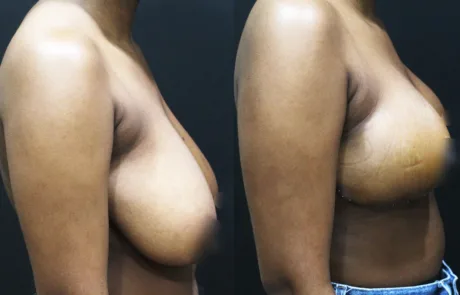
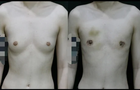
This patient had approximately B-cup breast volume prior to surgery. Because the breast size was relatively moderate, a keyhole or minimal-incision technique could have been considered as a surgical option.

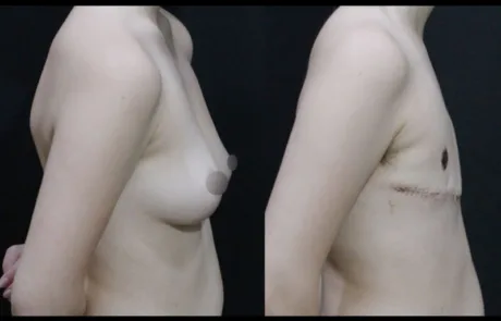
However, the patient clearly expressed a preference for achieving a completely flat masculine chest contour without any residual sagging or loose skin. For this reason, after careful consultation and surgical planning, we proceeded with a Double Incision Mastectomy with Free Nipple Graft, which is often the most reliable technique for achieving a flat chest in patients who want the most definitive contour correction.

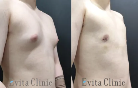
The photo shown here was taken two weeks after surgery, immediately following suture removal.

At this stage of recovery, the patient already shows minimal bruising and significantly reduced swelling, which indicates that the early healing process is progressing well.

Nipple Graft Healing Process

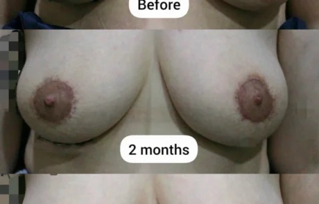
At two weeks after surgery, it is normal for the grafted nipple-areola complex to appear dark or covered with scab-like tissue. This is part of the expected healing process following a free nipple graft.

Over the next 3–4 months, the dark scab gradually separates and falls away. Underneath this layer, new pink tissue forms and the nipple-areola complex regenerates, eventually developing a more natural color and texture.

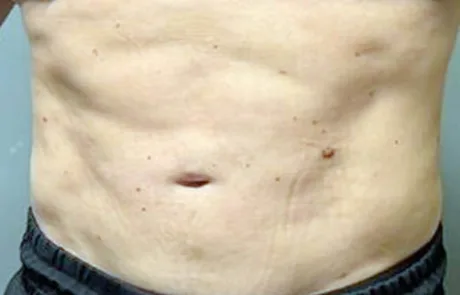
Chest Contour During Early Healing

At this early postoperative stage, some irregularity or slight contour unevenness can still be observed. There may also appear to be a subtle step-off between the upper chest skin and the lower abdominal skin.

This occurs because the skin thickness of the upper chest and lower torso is naturally different, and these tissues have been repositioned during surgery. As healing progresses and the tissues soften, this difference typically becomes much less noticeable.

Scar Healing and Management

The incision scars are still visible at this stage, which is expected only two weeks after surgery. Over time, these scars will gradually soften, flatten, and fade.

For optimal scar healing, patients are instructed to:

• Apply Steri-Strips or silicone tape for at least two months after surgery
• After the early healing phase, transition to topical scar management ointments or silicone gel

Consistent scar care is an important part of achieving the best long-term aesthetic outcome.

Return to Activity and Chest Development

After approximately one month, patients can gradually resume physical activity beginning with light stretching exercises.

As recovery progresses, strength training that targets the pectoralis muscles can help further enhance chest contour and definition. Developing the pectoral muscles often contributes to a more natural and masculine chest appearance after top surgery.

FTM top surgery before and after double incision mastectomy with free nipple graft at 2 weeks postoperative
breast lift side before after korea

Breast Lift (Anchor Mastopexy) with Superomedial Pedicle

2026-03-09T14:24:52+09:00

This patient had naturally full breasts and a relatively larger body frame. Her main concern was breast ptosis (sagging), but she strongly wished to maintain as much breast volume as possible after surgery.

To address her concerns, an Anchor Mastopexy technique was performed using a superomedial pedicle. This method allows for effective lifting and reshaping of the breast while preserving blood supply to the nipple-areola complex and maintaining as much natural breast volume as possible.

Before surgery, the patient was carefully informed that even when the primary goal is lifting rather than reduction, a slight decrease in apparent breast volume is often unavoidable. This occurs because excess skin must be removed to reposition the breast to a higher, more youthful position.

Another important consideration discussed with the patient was the long-term effect of gravity. In patients with naturally heavier breast tissue, the weight of the breast can gradually lead to some degree of recurrent sagging over time. In such cases, a secondary lifting procedure may be considered in the future if further correction is desired.

The postoperative result shows improved breast position, better contour, and a more youthful shape, while maintaining the patient’s overall breast volume and natural appearance as much as possible.

Breast lift before and after using anchor mastopexy with superomedial pedicle showing improved breast position and shape
breast lift side before after korea
Breast augmentation before and after side view using Mentor smooth round moderate plus 350cc implants at Evita Clinic Korea

Breast Augmentation with Mentor Smooth Round Moderate Plus 350cc

2026-03-06T17:25:50+09:00

Breast augmentation was performed using Mentor Smooth Round Moderate Plus implants (350 cc). The implants were placed using a dual-plane technique, through a 5 cm incision along the inframammary fold.

The surgical area was first anesthetized using the tumescent technique, and the procedure was performed under IV sedation.

The image shows the result 2 weeks after surgery. At this early stage, the pectoralis major muscle may still be slightly contracted as it adapts to the implant, which can temporarily make the breast appear somewhat elevated or tight.

Over the next 2–3 months, as the muscle gradually relaxes and the implant settles into position, the nipple position typically descends slightly and the breast contour becomes softer and more natural.

Breast augmentation before and after side view using Mentor smooth round moderate plus 350cc implants at Evita Clinic Korea
Areola reduction before surgery and postoperative results at 2 months and 4 months after circumareolar areola reduction surgery

Areola Reduction (Circumareolar Technique)

2026-03-06T17:27:03+09:00

Procedure: Areola Reduction
Technique: Circumareolar reduction
Anesthesia: Local anesthesia with IV sedation
Follow-up: 2 months / 4 months

Case Description

This patient presented with enlarged areolae that appeared disproportionate to the breast size. The goal of surgery was to reduce the diameter of the areola while maintaining a natural contour of the nipple-areola complex.

The procedure was performed using a circumareolar technique, in which a donut-shaped ring of pigmented skin is removed and the outer skin is gathered toward the smaller inner circle.

Some patients ask whether the areola can be reduced to less than 3 cm in diameter in a single procedure. However, this technique has anatomical limitations. When the outer circular skin edge is sutured to a much smaller inner circle, the difference in circumference becomes very large. According to the basic geometric relationship (circumference = 2πR), a large difference in radius creates a significant discrepancy in skin length. When this discrepancy becomes excessive, the surrounding skin must wrinkle to accommodate the difference.

This excessive tension may lead to:

  • visible wrinkling of the surrounding skin
  • more noticeable scars
  • slower scar maturation

In addition, if too much tension is applied, the nipple itself may become flattened due to the pulling force of the closure.

For this reason, it is important not to reduce the areola too aggressively in a single procedure. In cases of very large areolae, a staged reduction approach may be safer. The areola can be reduced partially first, allowing the skin to adapt, and then further reduced later if necessary. This approach generally produces a more natural shape and better scar quality.

In this patient, the areola appeared smaller at 2 months after surgery, but a slight enlargement was observed later as the skin tension gradually relaxed. This is a known characteristic of circumareolar reduction and must be considered during surgical planning.

Postoperative care is also very important. Patients are advised to maintain prolonged Steri-Strip support to help control scar tension and minimize the tendency for the areola to stretch again during the healing process.

Circumareolar areola reduction surgery showing enlarged areola before surgery and reduced areola after surgery at Evita Clinic
FTM top surgery before and 2 weeks after keyhole technique showing minimal scars and early healing stage

FTM Top Surgery (Keyhole Technique) – 2 Weeks After Surgery

2026-03-06T17:28:43+09:00

These photos show the results of an FTM top surgery performed using the keyhole technique, taken two weeks after surgery and immediately after suture removal.

This patient had very small breast tissue, which is the ideal condition for performing the surgery with a minimal incision. In cases like this, the keyhole technique allows us to remove the glandular tissue while leaving almost no visible scars on the chest.

In the postoperative photo, you may notice some bruising and small scabs around the suture removal sites. The chest may also appear slightly darker in some areas. This is a normal part of the healing process, and these signs typically improve quickly over the next few weeks as the bruising fades and the skin continues to heal.

Before surgery, the patient was also concerned about the size of the nipples. In most cases, when the breast gland is completely removed, the nipples naturally decrease in size by about 10–20%. In this case, the nipple diameter is approximately 1.2 cm, which is slightly larger than the average male nipple.

We will first observe how much the nipple size reduces naturally during healing. If the patient later wishes to make them smaller, a simple nipple reduction procedure can be performed.

The patient was very happy with the result and returned home with great satisfaction.

FTM top surgery before and 2 weeks after keyhole technique showing minimal scars and early healing stage
Gyne 202511 BNA

Gynecomastia, Grade 2, 20yrs

2026-03-06T17:31:20+09:00

Gyne 202511 1
Gyne 202511 2
Gyne 202511 3
Gyne 202511 4

Gynecomastia, Grade 2, 20 yrs old

After 2 week

This is a 20-year-old patient with gynecomastia.
He has a slim build, but the glandular tissue is relatively large, resulting in so-called “rocket-shaped” protruding breasts and a moderately enlarged areola.

Because he is young, we performed surgery using the keyhole technique with a minimal incision.
These photos were taken 2 weeks after surgery, before suture removal.

Since he is young, there is not much skin sagging at this stage.
However, at around 3 to 4 weeks, the sagging of the skin will appear more pronounced.
That said, after about 2 months, with regular massage and exercises to build the pectoralis major muscle, the skin laxity can be improved.

Even without performing a donut (periareolar) excision, we expect the areolar diameter to decrease by more than 20% as the skin contracts.

TOP nonbinary BNA

TOP Surgery, nonbinary, double incision

2025-01-10T14:42:29+09:00

TOP nonbinary B
TOP nonbinary A

TOP surgery, double incision without nipple graft

After 1 month

In the case of nonbinary individuals, if they do not wish to undergo free grafting of the areola and nipple during double incision surgery, the grafting is simply not performed.

As a result, the surgery is faster, and the recovery period is shorter.

The incision scars align with the natural lower border of the pectoral muscle, and with proper scar management, they can become less noticeable over time.

Go to Top