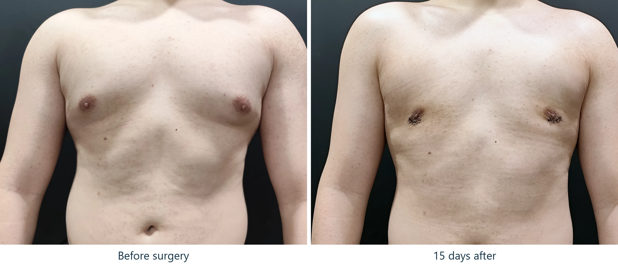
Gynecomastia Visible Only from the Side – 2 Weeks After Surgery
A chest that reads as nearly normal head-on, and unmistakably abnormal in profile.
- Concern
- Grade 2 gynecomastia, apparent in profile rather than head-on
- Procedure
- Liposuction with glandular excision through a periareolar incision
- Tissue excised
- 35 g on the left, with a comparable specimen on the right
- Photographs
- Before surgery and 15 days after
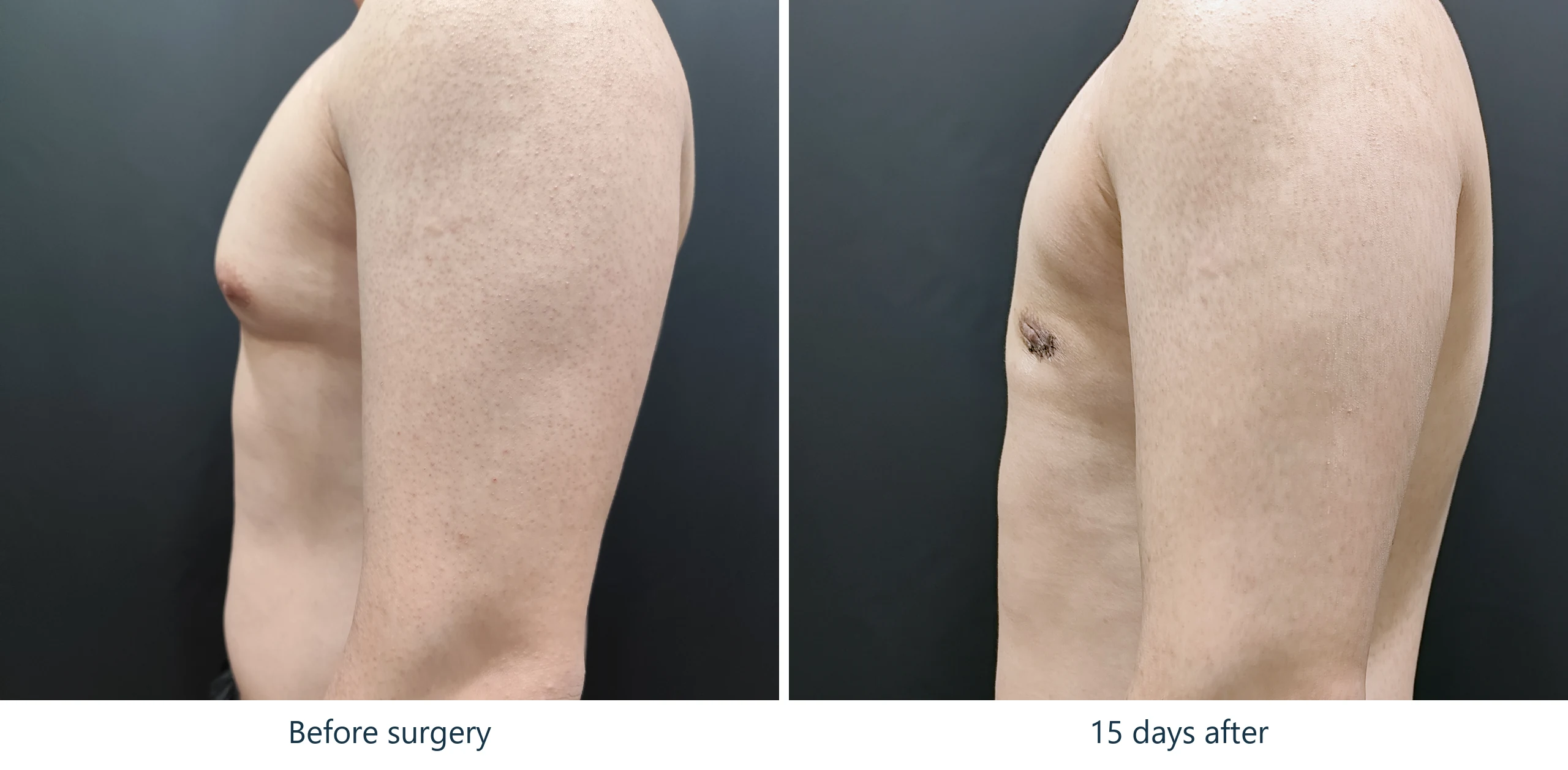
Gynecomastia is a problem of projection, and projection does not photograph from the front. Looking straight on, this chest is only slightly full and the nipples sit a little proud. Turn ninety degrees and there is a distinct cone of tissue with the nipple at its tip. Both photographs are of the same chest on the same morning.
Why the front view understates it
A frontal photograph flattens the chest into an outline. It shows width and it shows the areola, but it cannot show how far the tissue stands away from the ribs — and that distance is the whole complaint.
This is why patients with exactly this pattern are so often told they are imagining it. Friends and family look at them head-on, see a chest that is more or less flat, and say so. The patient, meanwhile, is looking down at themselves, or catching a side profile in a changing-room mirror, and seeing something quite different. They are both looking at the same chest from different angles, and only one of those angles carries the information.
If you are unsure whether what you have is gynecomastia, photograph yourself from the side with your arm down, not from the front. The side view is the one that answers the question.
Evita Clinic
What the photographs show
Before surgery
In profile, firm tissue lifts the lower chest into a rounded cone and carries the nipple forward and slightly downward. Head-on, the same chest shows only mild fullness with somewhat prominent areolae. The dominant tissue here is glandular, which is why it holds a defined shape rather than draping the way fat does.
15 days after surgery
The profile is flat: the cone is gone and the chest wall runs in a straight line from the collarbone down to the abdomen. The periareolar incisions are visible as dark crusted lines at the lower edge of each areola, still healing rather than yet scars. Bruising has largely cleared by this point.
Why liposuction alone would not have worked
Glandular tissue is dense and fibrous and will not pass through a liposuction cannula. Removing only the surrounding fat leaves the firm disc in place, and with the softer tissue around it gone, that disc often becomes more conspicuous rather than less. The gland has to be excised, and excision requires an incision.
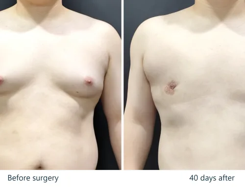
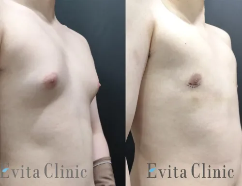
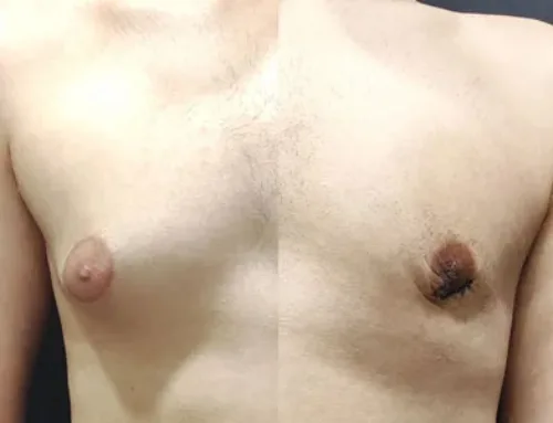
We have set out that trade-off in more detail, along with what the first six weeks of recovery actually look like, in a separate case photographed at 8, 15 and 40 days.
The scar
The incision follows the lower border of the areola, where pigmented skin meets ordinary chest skin. That colour change disguises the line well, but the scar is permanent. At two weeks it is still a healing wound; scars of this kind continue to fade over roughly 6 to 12 months.
What this operation did not do
The abdomen is unchanged between the two photographs, and was never the target. Gynecomastia surgery corrects the chest. It is not a weight-loss operation and it does not contour the trunk.
Two weeks is early. These are the earliest photographs we would publish, and they are not a settled result. Swelling is still present in the operated area at this stage, the incisions are healing wounds rather than mature scars, and the final contour will continue to refine over the following weeks and months. An early photograph misrepresents this operation in both directions — the chest is still slightly swollen, and the scar looks angrier than it will.
- Gynecomastia is a problem of projection, which a frontal photograph cannot show.
- A chest can look nearly flat head-on and still carry a clear glandular cone in profile.
- If you are assessing yourself, photograph from the side with the arm down.
- Glandular tissue cannot be suctioned; it has to be excised, and that leaves a permanent areolar-border scar.
- These photographs are at 15 days and are not a settled result.
About these images
The two sessions were photographed at different camera distances. The post-operative frames have been scaled so that the shoulder and collarbone silhouette matches the pre-operative frame — a region this operation does not alter — with the alignment solved by computer rather than by eye, and the same crop applied to both panels. Brightness and colour were matched between sessions using the backdrop and the shoulder skin as references.
Nothing has been stretched, slimmed, reshaped or retouched. Identifying features have been cropped out of frame rather than painted over.
Results vary depending on individual anatomy, healing response, lifestyle factors and postoperative care. These images are provided for informational purposes only and do not guarantee specific results.