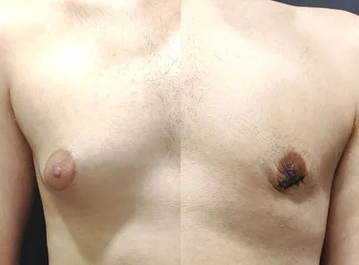
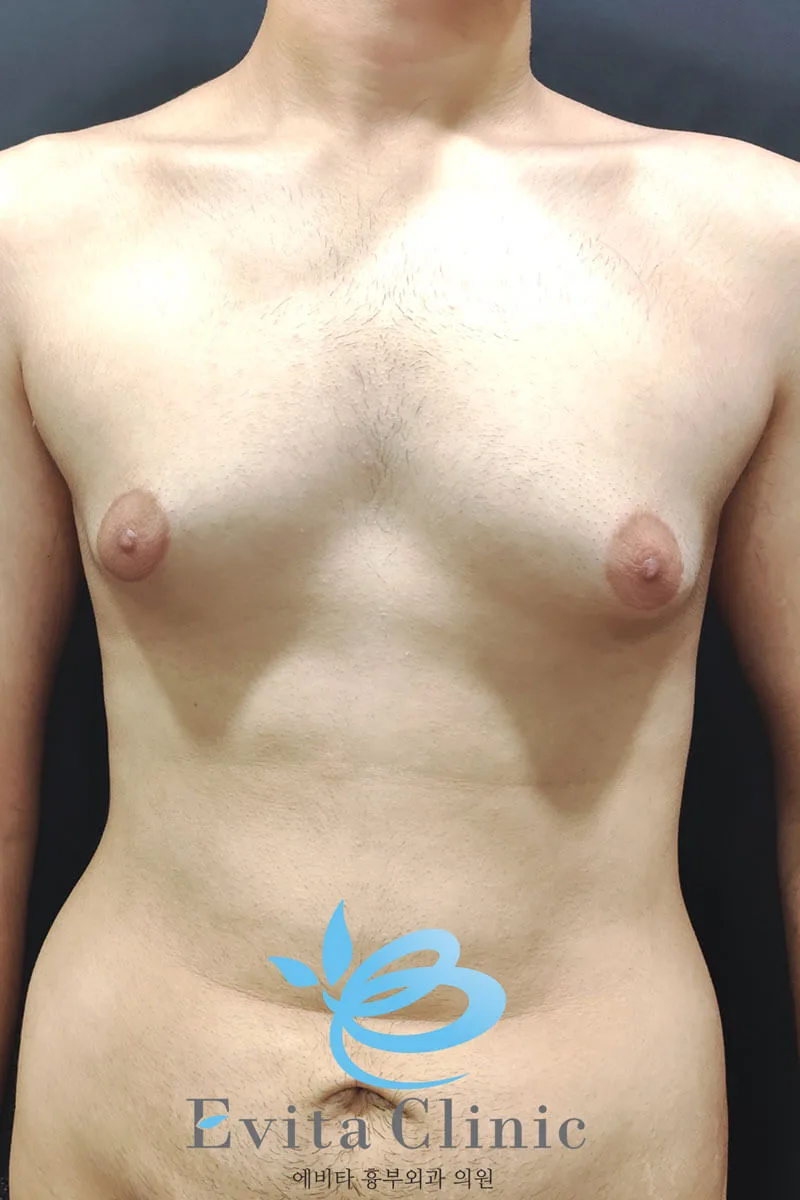
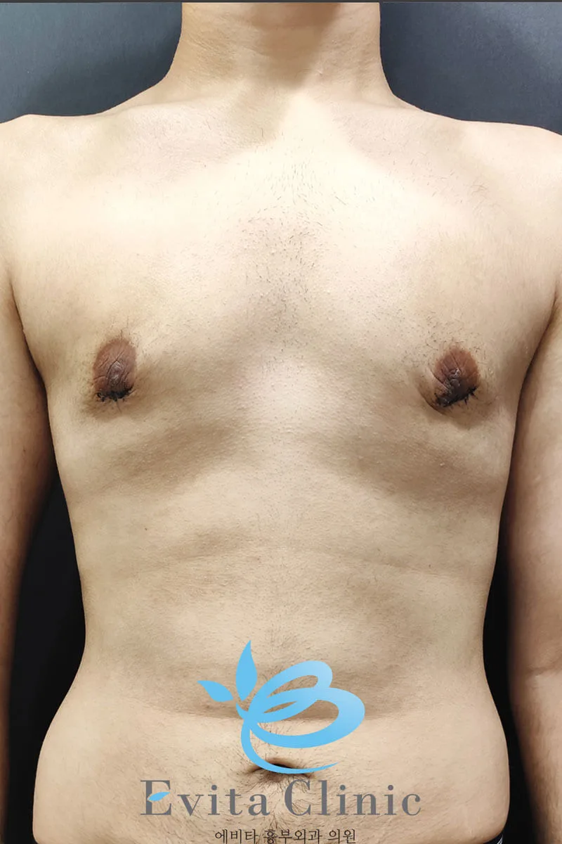
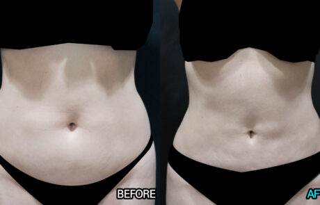
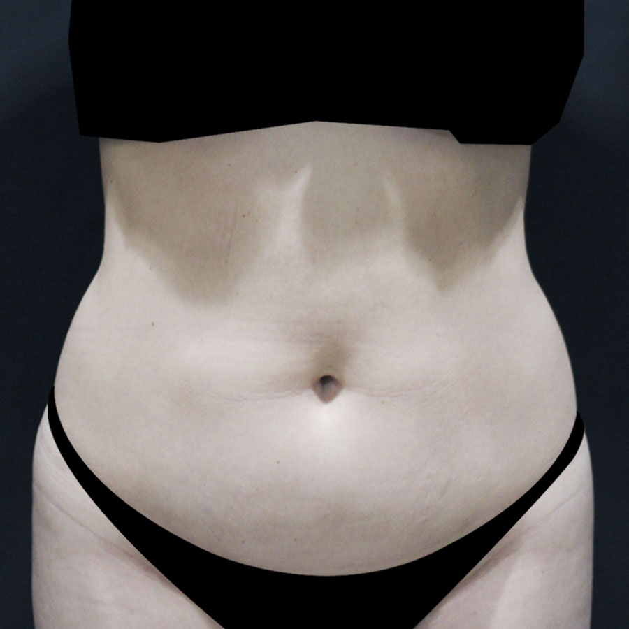
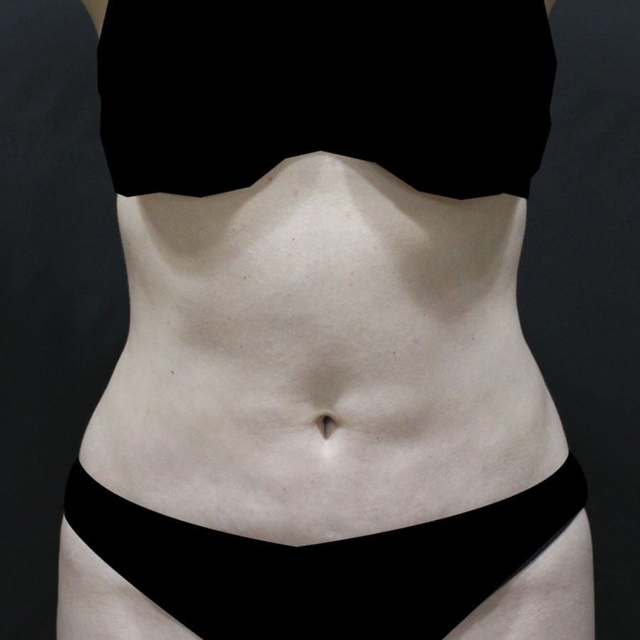
Gynecomastia (Grade 2) – 4 Weeks After Surgery
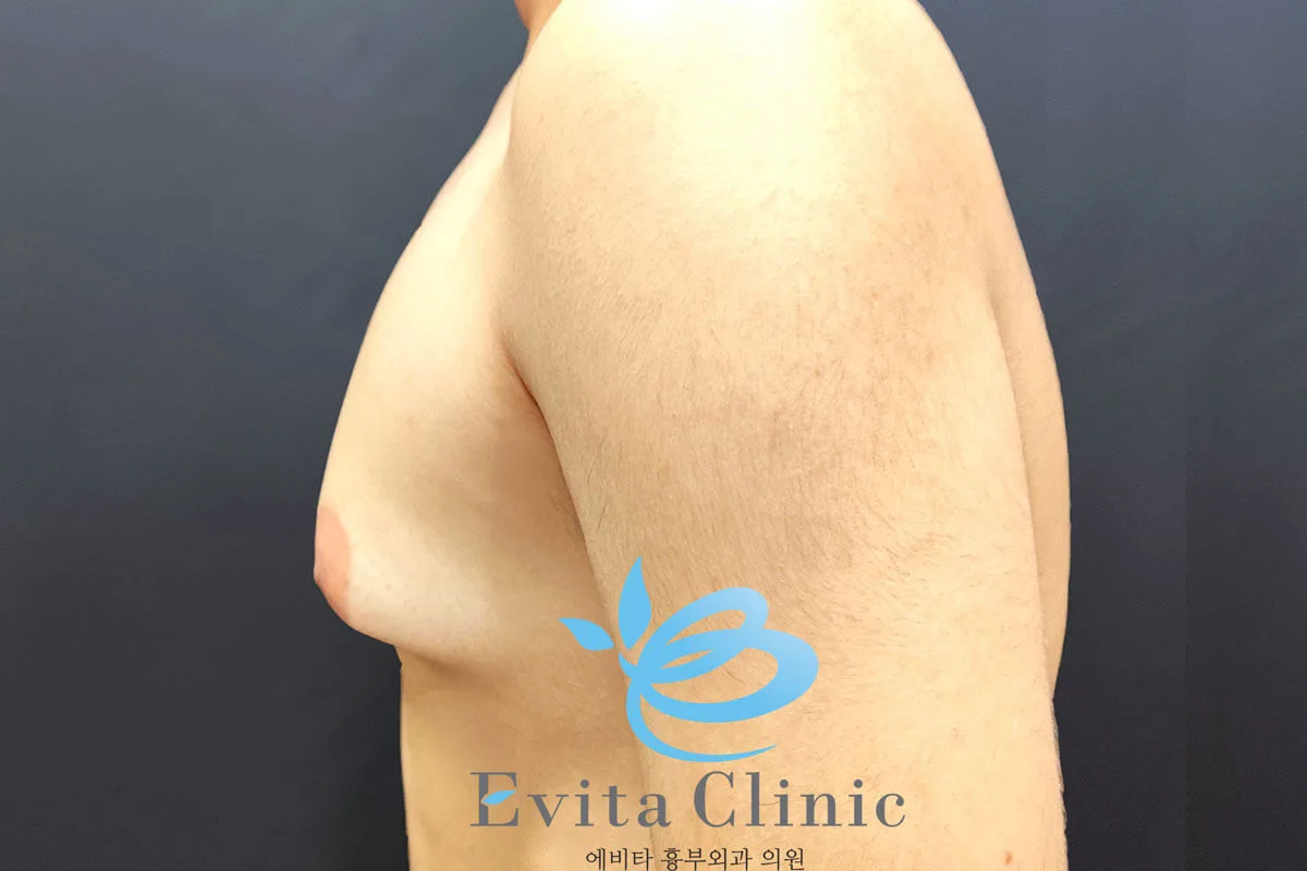
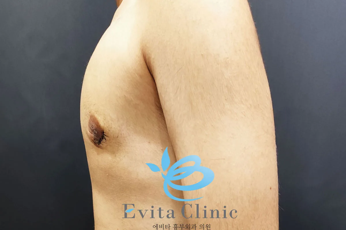
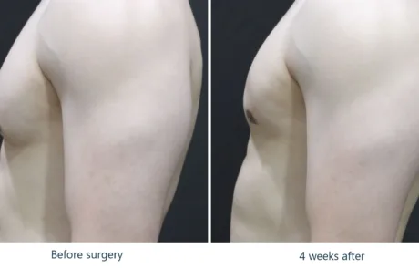
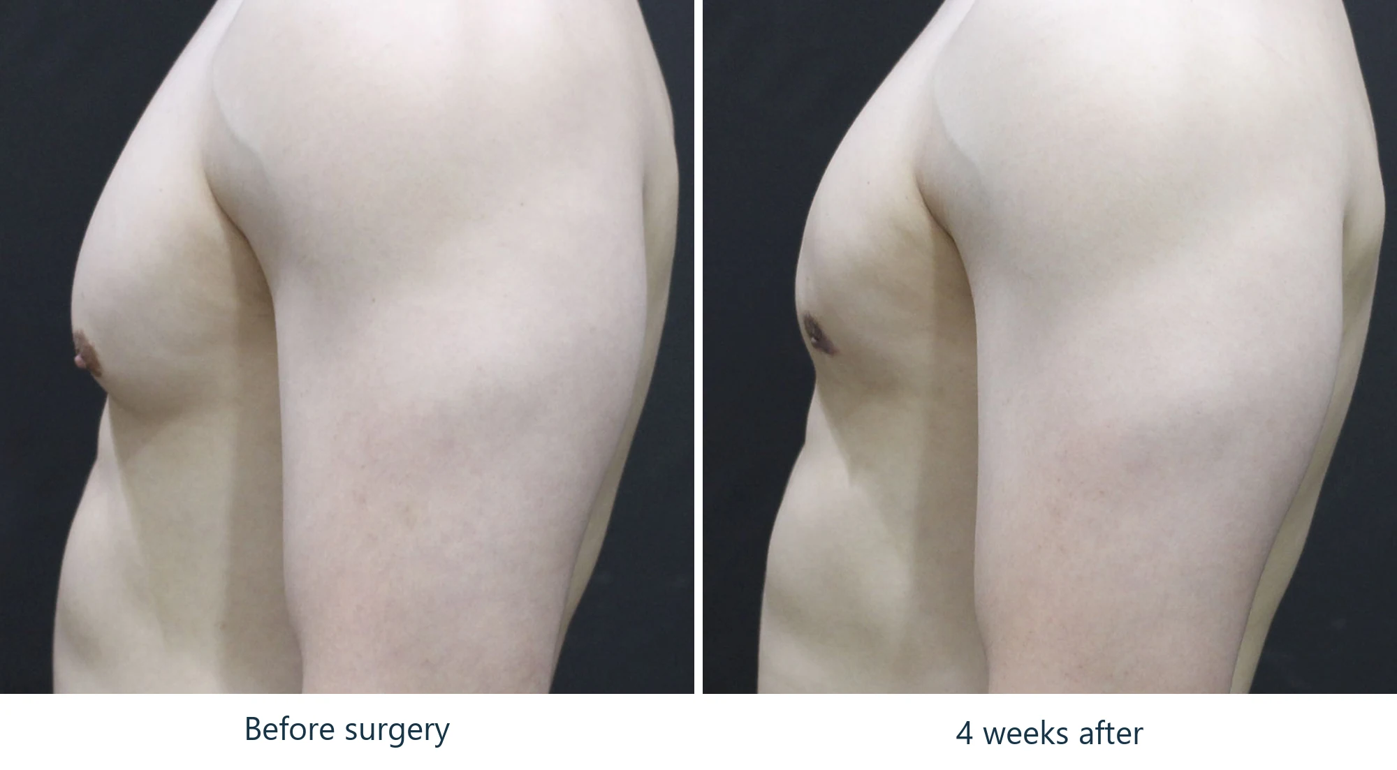
Seen from the side, where the shape of gynecomastia actually lives.
GynecomastiaGrade 24 weeks post-op
- Concern
- Grade 2 gynecomastia — a firm cone behind the nipple
- View
- Lateral (side profile)
- Photographs
- Before surgery and 4 weeks after
Gynecomastia is a problem of projection, and projection only shows from the side. Before surgery this chest rises into a defined cone with the nipple carried forward at its tip. At four weeks the profile runs straight from the collarbone down to the abdomen. That is the change the operation makes, and it is the change a frontal photograph tends to hide.
Why the side view is the one that matters
A frontal photograph flattens the chest into an outline. It shows width and it shows the areola, but it cannot show how far the tissue stands away from the ribs — and that distance is the whole complaint. Turn ninety degrees and the same chest tells you immediately what grade it is.
If you are unsure whether what you have is gynecomastia, photograph yourself from the side with your arm down. That is the view that answers the question.
What changes, and when
The contour is corrected at surgery. The cone behind the nipple is removed and does not come back. What takes time is everything else: swelling, firmness, and the scar at the areolar border.
Four weeks is far enough along that the shape is readable and most of the bruising has gone. It is not far enough along to call the result settled — deep swelling continues to resolve for some months, and the scar goes on maturing for 6 to 12.
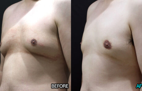
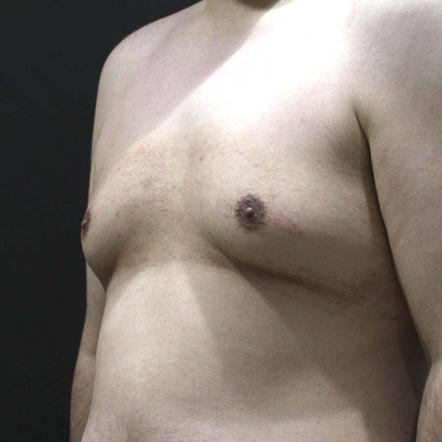
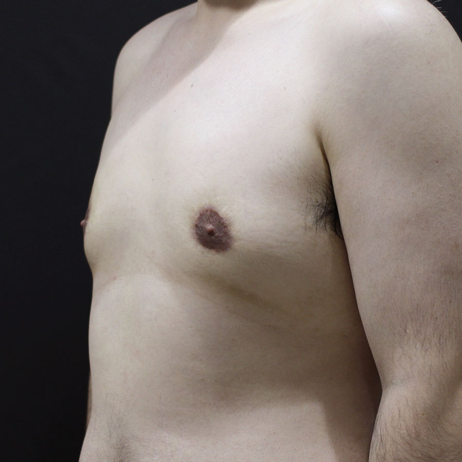
Four weeks is an intermediate point, not a final result. The chest is still slightly swollen at this stage and the scar has not begun to fade. Judging gynecomastia surgery from a one-month photograph understates it in both directions — the contour will refine further, and the scar will become far less noticeable than it is here.
- Gynecomastia is judged in profile, not head-on.
- The cone behind the nipple is glandular tissue; removing it is the operation.
- Contour is corrected at surgery; swelling and scar take months.
- Four weeks shows the shape, not the finished result.
About these images
These are clinical photographs from this patient’s own records, published with consent. Identifying features are outside the frame.
The two frames have not been geometrically normalised. They were taken in separate sessions at slightly different camera distances, and we have not rescaled either one to compensate. Nothing has been stretched, slimmed, reshaped or retouched; the only change is that the two original photographs have been placed side by side and labelled.
Results vary depending on individual anatomy, healing response, lifestyle factors and postoperative care. These images are provided for informational purposes only and do not guarantee specific results.